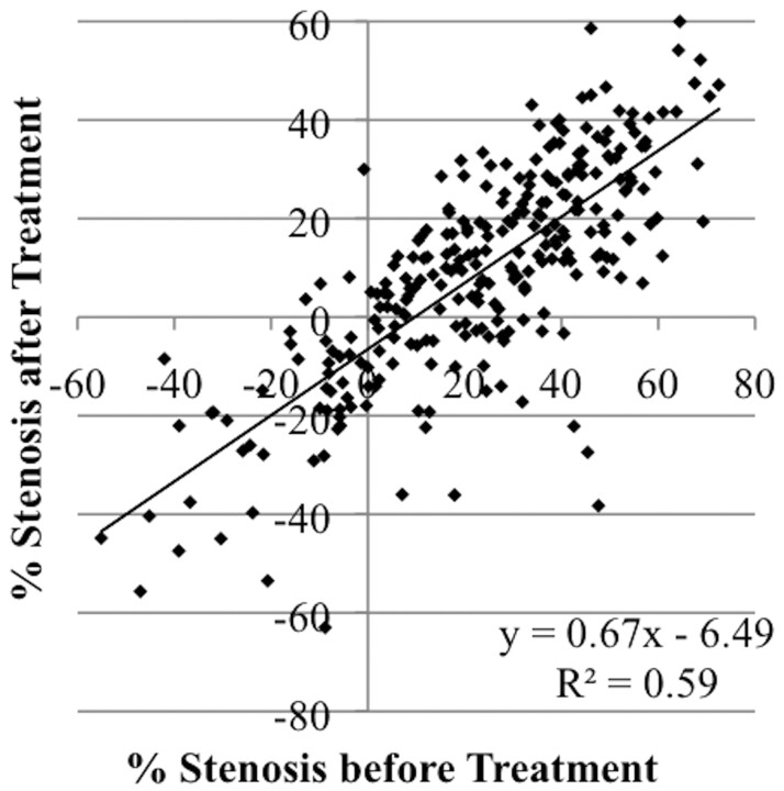
Fig 2.
Scatterplot demonstrating improvement in angiographic cerebral vasospasm after treatment with intra-arterial verapamil. During 16 procedures in 12 patients, 34 arterial distributions were selectively treated. Cerebral angiograms were obtained after patient presentation with SAH and days later before and after intra-arterial verapamil treatment of cerebral vasospasm. Artery diameters were measured in a blinded fashion at multiple predetermined sites (whether or not vasospasm was present) in each arterial distribution from each of the 3 angiograms, and the resulting 275 data points were plotted.

