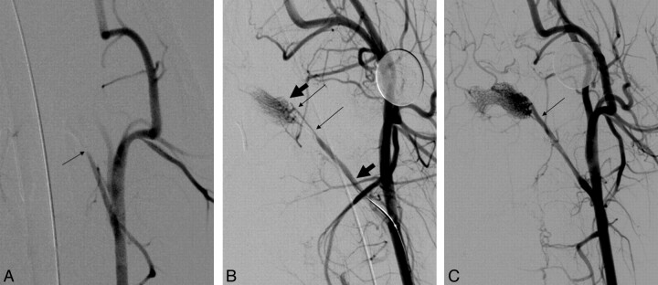
Fig 2.
A, Anteroposterior angiogram of the left common carotid artery in swine. Occlusion of the APA is seen (arrow). B, Right anterior oblique angiogram of the left common carotid artery with the device deployed shows immediate flow restoration. The thin arrows indicate the proximal and distal aspects of the clot, while the thick arrows point to the proximal and distal ends of the device. C, Right anterior oblique angiogram of the left common carotid artery postretrieval of the clot shows complete restoration of flow. Arrow shows the original site of occlusion.

