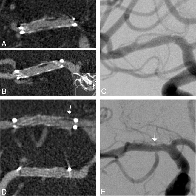
Fig 1.
Follow-up images of a 56-year-old woman 7 months after ICAS of a 76% middle cerebral artery stenosis. A and B, Excellent delineation of in-stent anatomy on coronal (A) and curved planar (B) ivACT reformations allows exclusion of a high-grade ISR and detection of low-grade neointimal hyperplasia within the body of the stent. C, The same findings are demonstrated on the iaDSA image. D and E, Another example of a low-grade restenotic lesion (25%), which is depicted in the proximal stent portion (arrows) on both ivACT (D) and a standard iaDSA image (E). This 61-year-old woman was admitted to standard follow-up without any new symptoms after ICAS of a symptomatic 79% middle cerebral artery stenosis. At the confines of the Wingspan stent, streak artifacts limit the image quality on ivACT reformations.

