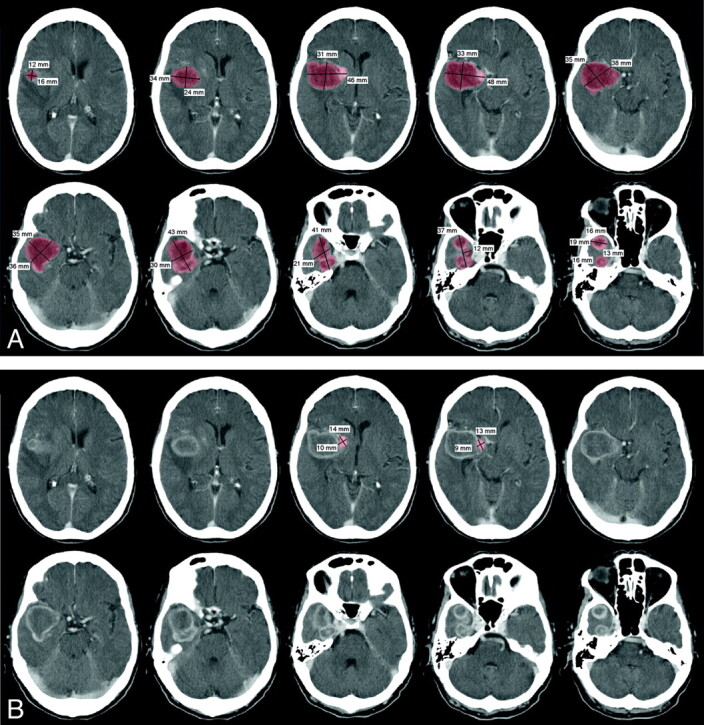
Fig 2.

CT brain scan with intravenous iodinated contrast of a patient with a partially thrombosed, giant right middle cerebral aneurysm. A, 5-mm contiguous axial images through the aneurysm demonstrate a ring-enhancing mass with a maximal dimension of 4.8 cm and a total volume of 48 mL. B, The patent component of the aneurysm measures only 14 mm in maximal dimension with a volume of 1.33 mL. The opacified component of the aneurysm at catheter arteriography represents 2.8% of the total aneurysm volume.
