Figure 1.

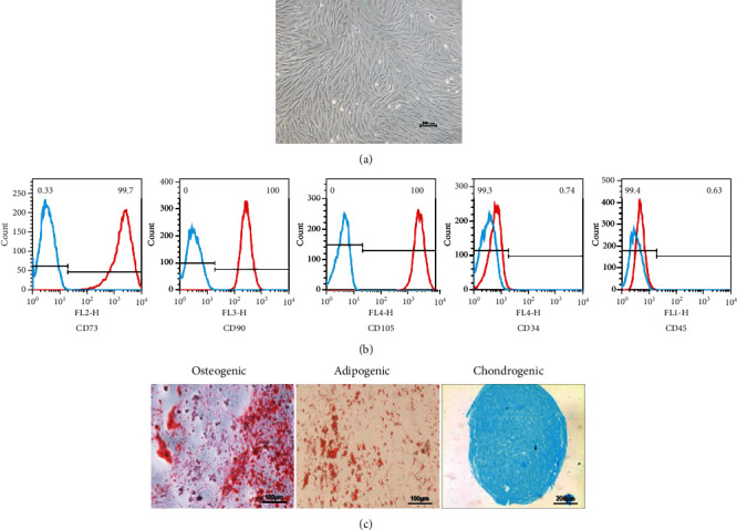
Isolation and identification of IPFP-MSC. (a) The P3 generation IPFP-MSC. (b) The results of flow cytometry. Blue line: negative control; red line: IPFP-MSCs. (c) Three-line differentiation experiments.

Isolation and identification of IPFP-MSC. (a) The P3 generation IPFP-MSC. (b) The results of flow cytometry. Blue line: negative control; red line: IPFP-MSCs. (c) Three-line differentiation experiments.