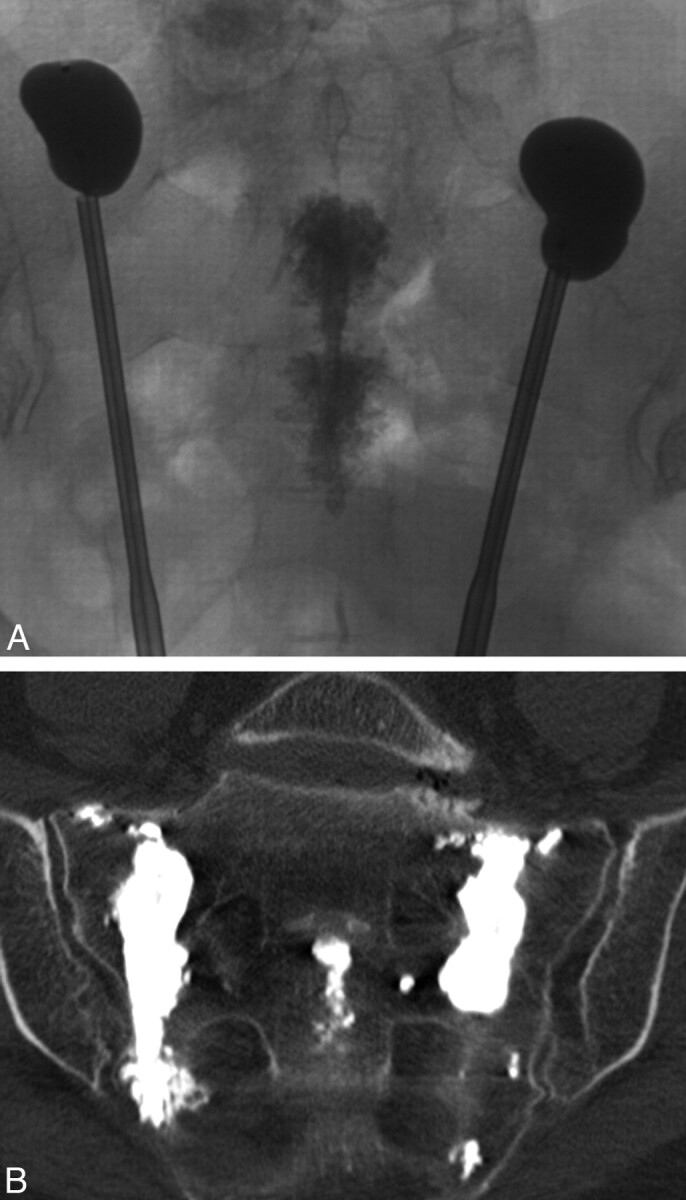
Fig 10.

Intraoperative radiograph (A) and postprocedural coronal oblique CT scan (B) from long-axis-approach sacral kyphoplasty demonstrate more even deposition of PMMA cement along the lateral aspect of the sacrum. Note the cement within the sacral bodies from midline-approach sacroplasty, performed immediately prior. Courtesy of Frank R. Hellinger, Orlando General Hospital, Orlando, Florida.
