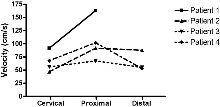
Fig 3.
Maximal BFV in centimeters per second measured with the ComboWire in the ICA at the cervical level (Cervical), proximal to the aneurysm (Proximal), and distal to the aneurysm (Distal). Intra-aneurysmal measurements are not plotted here because we considered them to be less accurate (see “Data Analysis”). Maximal BFV was calculated by averaging 5 consecutive peaks of the ComboMap flow signal. One measurement is lacking because the wire could not be placed distal to the aneurysm in patient 1 due to unfavorable geometry.

