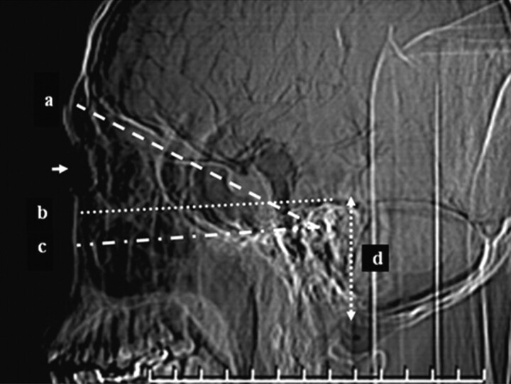
Fig 1.
Scanning baseline and coverage of temporal bone CT: glabellomeatal line, the baseline of the sequential axial scanning mode (a); a line parallel to the acanthiomeatal line passing by the superior edge of the temporal bone, the upper limit of the modified helical scanning mode (b); the acanthiomeatal line approximately parallel to the hard palate (c); and the scanning range of the temporal bone CT (d). The arrow is the position of lens.

