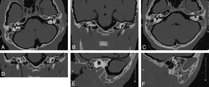
Fig 2.
Sequential axial and coronal CT images and reformatted images from the modified helical scan. A and B, Sequential axial section of the vestibulum auris (A) and a coronal section of the cochlea (B), with the structures delineated vividly. C–F, Axial, coronal, and sagittal reformatted sections from the modified helical scan with the microanatomy displayed clearly and more diagnostic information delivered.

