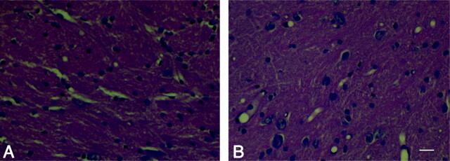
Fig 4.
Pathologic change after irradiation. A and B represent the results of HE staining in the control group (A) and the 40-Gy irradiated group 20 days after irradiation (B). It was noted that the partial loose and irregular arrangement of neurons and vacular degeneration in the parietal white matter near the cortex were found in the 40-Gy irradiated group 20 days after irradiation. Scale bar, 40 μmol/L.

