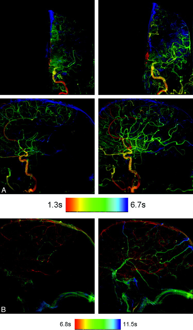
Fig 2.

A, AP and lateral color-coded images obtained before (left column) and after (right column) angioplasty and stent placement of a left middle cerebral artery stenosis. The posttreatment images clearly show faster arterial filling with greater opacification of the middle cerebral artery distribution. B, Color-coded images of lateral projections of the left internal carotid angiogram obtained before (left image) and after (right image) angioplasty and stent placement. Notice the greater extent of parenchyma opacification and more normal filling on the posttreatment image.
