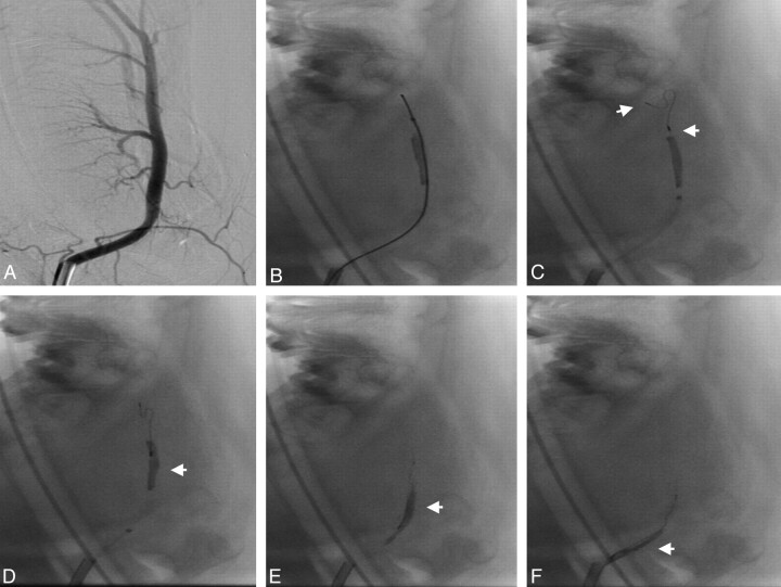
Fig 2.
Angiogram of the lingual artery before embolization (A). Passing of the microcatheter and microwire between thrombus and the vessel wall (B). Unsheathing of the Phenox CRC distally to the thrombus (C; proximal and distal markers: arrows). Retrieval of the device (D–F). The thrombus is mobilized in a stretched position (arrows) by the device during retrieval without compression or compaction.

