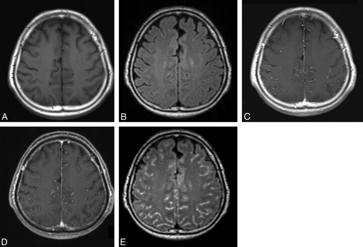
Fig 2.
MR images in a 66-year-old man (case 7) with viral meningitis treated with an antiviral agent. A and B, Precontrast T1-weighted (A) and 3D T2-FLAIR (B) images show no apparent leptomeningeal abnormality. C and D, Postcontrast T1-weighted (C) and MPRAGE (D) images depict enhancement in the sulci, corresponding to the vessels. Compared with the postcontrast T1-weighted image (C), the postcontrast MPRAGE image does not provide additional information. Both reviewers ranked the MPRAGE sequence as grade 0. E, Postcontrast 3D T2-FLAIR image shows abnormal leptomeningeal enhancement in the sulci. Compared with the postcontrast T1-weighted image (C), postcontrast 3D T2-FLAIR provided additional information about abnormal leptomeningeal enhancement. Both reviewers scored the 3D T2-FLAIR sequence as grade 3.

