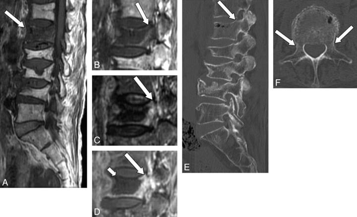
Fig 3.
A 76-year-old woman with benign compression fracture, which occurred 1 month earlier. A and B, T1WI shows diffuse hypointensity in the vertebral body and in the left pedicle of L1 (arrow). C, STIR image shows heterogeneous hyperintensity in these areas (arrow). D, Contrast-enhanced T1WI shows marked enhancement (arrow). A large area of low signal intensity indicates necrosis or cleft (small arrow). E and F, On sagittal (E) and axial (F) CT scans, there is no apparent abnormality in the pedicle (arrow).

