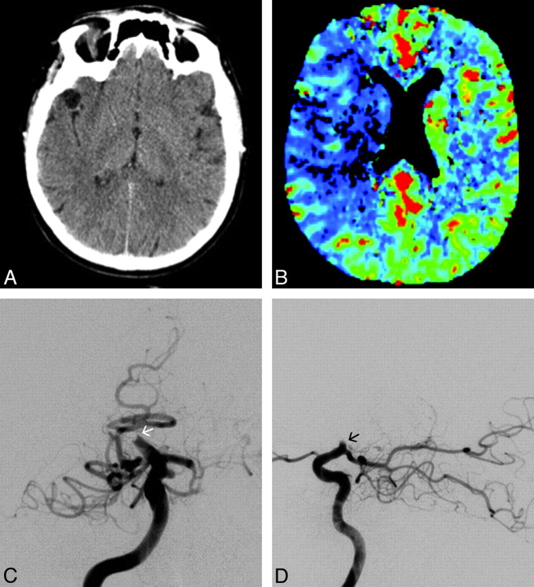
Fig 1.

Diagnostic illustration of a patient with acute stroke before treatment with the PS. A and B, Axial unenhanced CT scan (A) and CT perfusion cerebral blood flow map (B) obtained 2.5 hours after symptom onset demonstrate only subtle changes in the brain but a significant perfusion deficit in the right MCA territory. C and D, Selective right ICA injection angiogram, posteroanterior (C) and lateral (D) views, shows complete thromboembolic occlusion (arrows) of the ICA at the level of the origin of the anterior choroidal artery, which is still filling.
