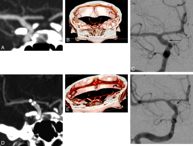
Fig 7.
Right supraclinoid ICA blister aneurysm, pre- and posttreatment. The blister aneurysm is not detectable on initial CTA images (A and B) but is clearly delineated on the correlative DSA image (black arrow, C). On the short-term posttreatment CTAs (D and E), the residual aneurysm remains invisible, while it is still clearly seen on the correlative posttreatment DSA image (black arrow, F).

