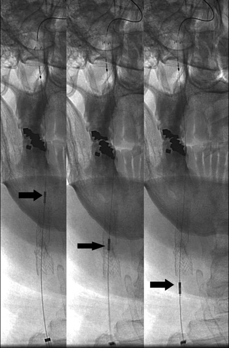
Fig 4.

Angiographic images demonstrate a retraction of the intravascular sonographic probe (arrows) by using a motorized device (not shown) after carotid stent placement to visualize any residual stenosis and apposition of the stent to the surrounding vessel wall.
