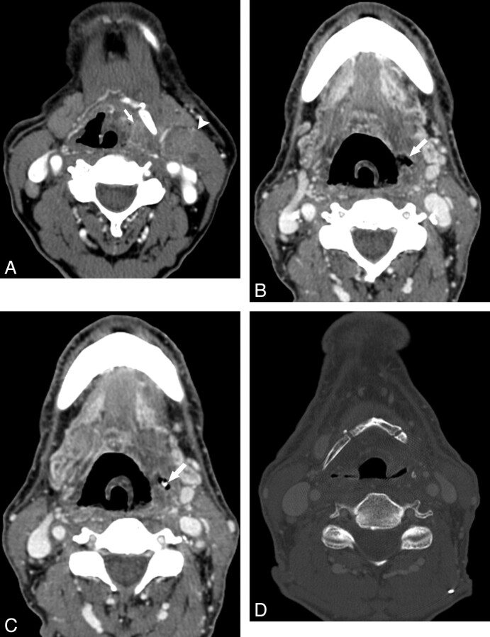
Fig 1.
Patient 4. A 64-year-old man radiated for squamous cell carcinoma of the left lateral oropharyngeal wall. A, CT examination with contrast obtained before radiation therapy shows the lower aspect of the lesion in the vallecula, lying adjacent to the hyoid bone (arrow). There is a nodal metastasis in the left neck (arrowhead), also adjacent to the hyoid. B and C, Axial contrast-enhanced CT images obtained 5 months following treatment with 70 Gy show a defect in the lateral pharyngeal wall (arrow, B) and an exposed piece of hyoid (arrow in C). At this time, the hyoid was otherwise intact. D, CT bone window on follow-up imaging shows a missing resorbed hyoid.

