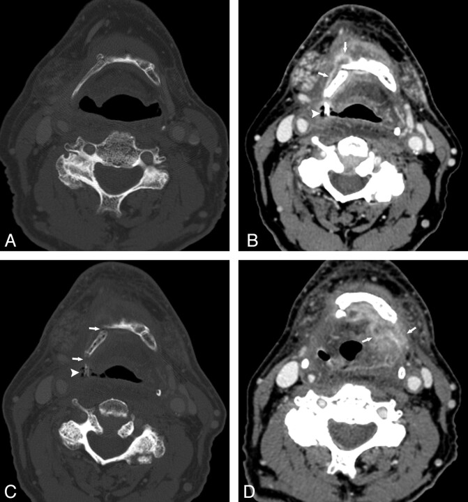
Fig 4.
Patient 3. A 66-year-old man status post 60 Gy for treatment of metastatic squamous cell carcinoma to the left neck, primary unknown, who subsequently developed a primary malignancy in the right oropharyngeal wall, for which an additional dose of 50.2 Gy was administered. A, CT bone window before XRT shows an intact hyoid. B and C, Contrast-enhanced CT, soft-tissue and bone windows, respectively, 5 months following completion of the second course of XRT. There is an enhancing process surrounding the right side of the hyoid (arrows, B) and an exposed fragment of hyoid surrounded by air (arrowhead, B and C). Hyoid fractures can be seen (arrow in C). D, Contrast-enhanced CT image 5 months after B and C shows dramatically increased enhancement extending to the left of midline (arrows). Because of these findings and relentless aspiration, a total laryngectomy was performed; at histologic examination, there was only necrosis and inflammation.

