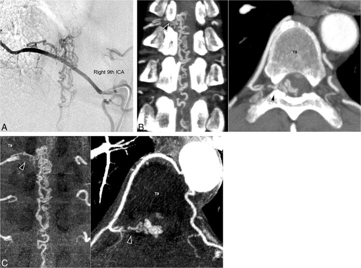
Fig 4.
Images of an illustrative case of a 53-year-old man (case 5). Spinal digital subtraction angiography of the right ninth ICA (A) shows a feeder and enlarged vein. C-CTA image (B) and R-CTA (C) at the T9 level can detect the feeder (black arrowhead) from the right ninth ICA. In R-CTA with thin-slab maximum-intensity-projection images, assessing the feeder and continuity is easier than with C-CTA. The window level and width of R-CTA are set to 120 and 240, respectively.

