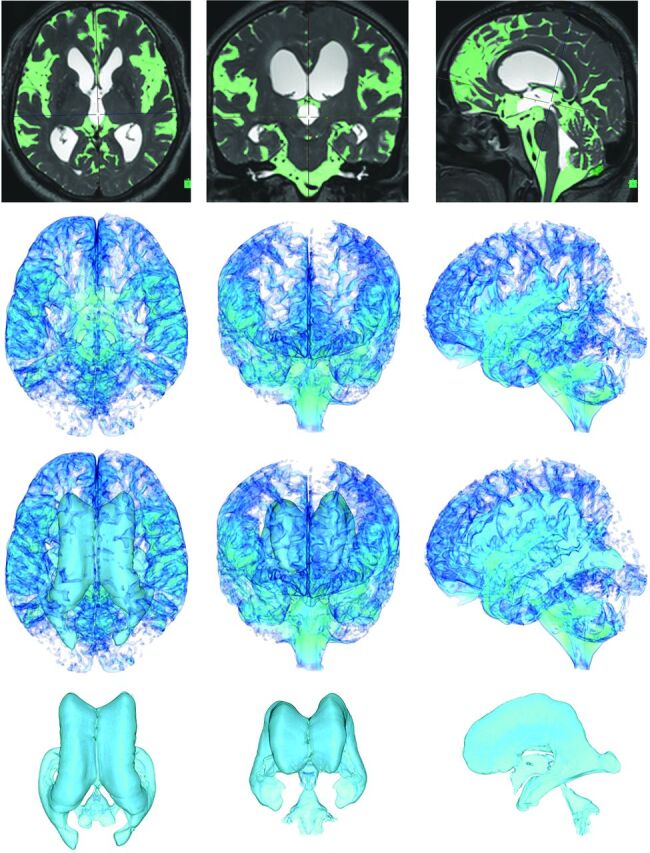
Fig 1.

Automatic extraction of CSF space. The figures in the top row show the MIP images on the T2-weighted 3D-SPACE sequence in the representative iNPH case. Light green indicates the subarachnoid space segmented automatically at a threshold intensity of >700 on the SYNAPSE 3D workstation. The other figures show the 3D volume-rendering reconstruction images of the subarachnoid space on the second line, total CSF on the third line, and ventricles on the last line. The left, middle, and right column figures show axial, coronal, and sagittal dimensional views, respectively.
