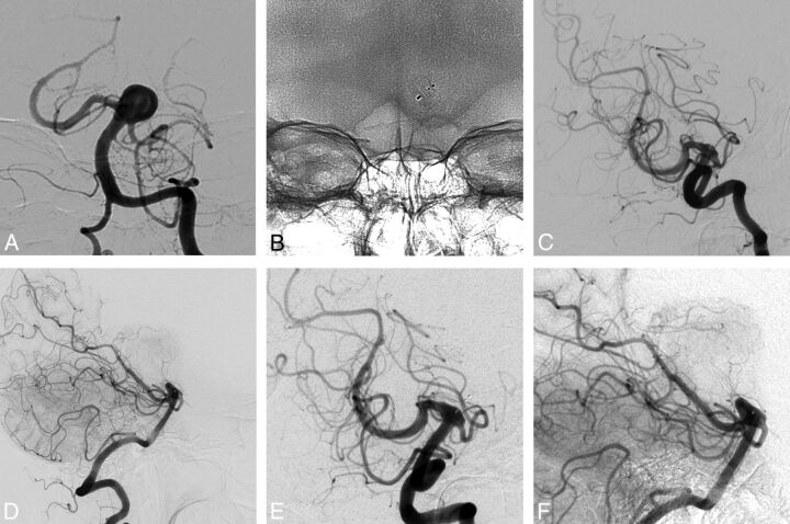
Fig 3.
A, Preoperative DSA shows a wide-neck basilar artery aneurysm. B, The WEB device is deployed in the aneurysm. C and D, Six-month DSA (oblique and lateral views) shows a small aneurysm remnant. E and F, Twenty-one-month DSA (oblique and lateral views) shows that the aneurysm remnant has not grown.

