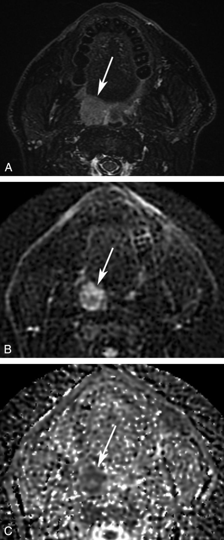
Fig 1.

Axial MR images of an HPV-positive OPSCC. A 48-year-old patient was diagnosed with an OPSCC in the right palatine tonsil. A, STIR MR image shows a hyperintense mass in the palatine tonsil (arrow). A b=750 diffusion-weighted image (B) and an ADC map (C) show decreased ADC. An ROI was drawn on the b=750 diffusion-weighted image and copied to the ADC map. In this ROI, ADCminimum measured 0.992 × 10⁻³ mm2/s.
