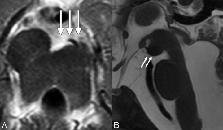
Fig 5.
A, Axial T2-weighted MR image shows asymmetry of the cerebral peduncles, giving them an appearance of a decaying molar tooth on axial images (white arrows). B, Midsagittal T2-weighted MR image reveals a dysmorphic mesencephalon (white arrows) and an enlarged prepontine cistern with increased vertical orientation of the brain stem.

