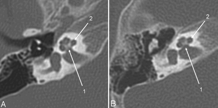
Fig 2.
Axial CT section of the right temporal bone obtained with a CTDIvol of 63 mGy (A) (14-month-old patient; DLP, 223 mGy cm; estimated Deff, 1.4 mSv) and with the low-dose protocol (B) (16-month-old child; CTDIvol, 10.8 mGy; DLP, 46.9 mGy cm; estimated Deff, 0.35 mSv). Critical structures like the modiolus and the thin bony lamella separating the internal auditory canal from the cochlea (1) and the spiral osseous lamina (2) are delineated well despite the higher image noise.

