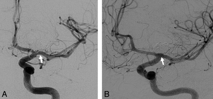
Fig 2.
Serial angiographic images of a 48-year-old man who underwent endovascular treatment for a ruptured left ICA terminus aneurysm. A, Immediate postprocedural image demonstrates a grade I protrusion. B, Six-month follow-up corresponding image demonstrates spontaneous resolution of the coil protrusion.

