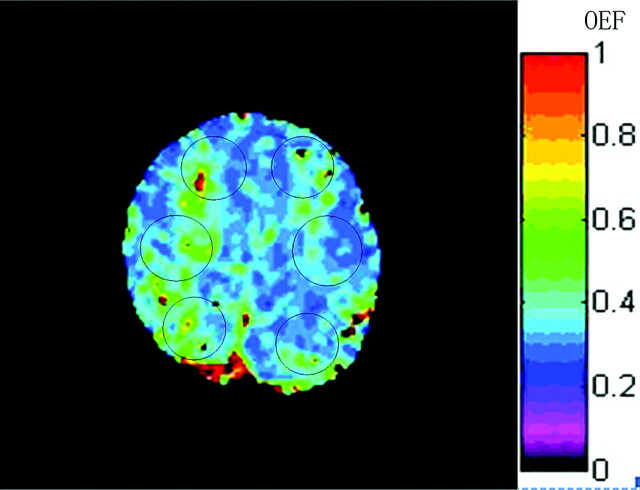
Fig 1.
Representative region-of-interest placement for OEF measurement in the study, shown on the OEF map from a patient. Each region of interest was placed in the anterior, middle, and posterior parts of bilateral hemispheres, which correspond to the territory of the ACA, MCA, and borderzone of the MCA and posterior cerebral artery, respectively. CBF was measured in the same way.

