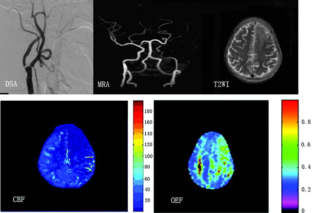
Fig 4.
A patient with territory infarction 2 weeks earlier due to left ICA severe stenosis. DSA and MRA show severe stenosis of the left ICA and no visualization of the bilateral ACAs and left MCA. T2-weighted MR image shows a left frontal infarction (top right). Severe reduction of CBF is noted in the anterior and middle parts of the left hemisphere (bottom left), which shows a markedly elevated OEF (bottom right).

