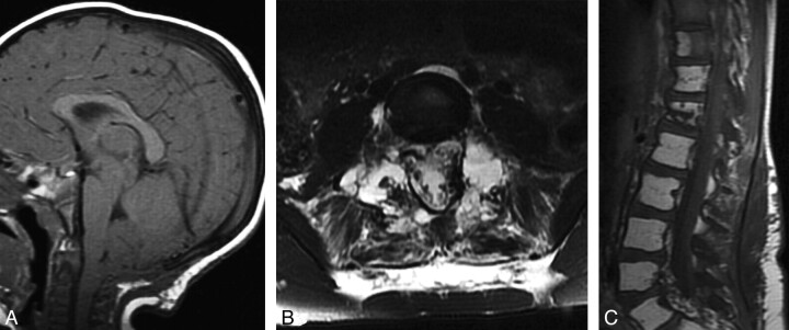
Fig 1.
A, Sagittal T1-weighted MR image obtained at 22 months of age after suboccipital decompression shows persistent cerebellar tonsillar ectopia and effacement of the prepontine cistern due to posterior fossa sagging. B and C, Axial T2-weighted (B) and sagittal T1-weighted (C) spine MR imaging at 7 years of age show extensive replacement of osseous structures by fluid-filled cystic spaces (B) and fatty marrow replacement in multiple vertebrae (C). Also, note fluid signal intensity in the paraspinous muscles and surrounding soft tissues (B).

