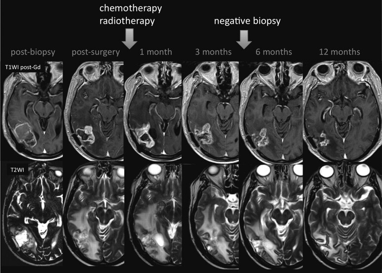
Fig 1.
Pseudoprogression. A 59-year-old man with GBM. An MR image obtained 1 month after RT-TMZ demonstrates an expansion of the right temporal lesion. Reductions in both the enhancing portion and the surrounding abnormal hyperintense area in the T2-weighted imaging were seen in the follow-up MR imaging examinations

