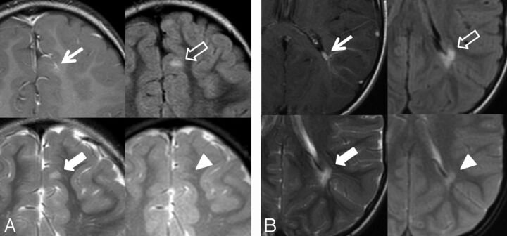
Fig 1.
Two DVAs with associated signal abnormality in a 5-year-old boy (A) and a 7-year-old boy (B). T1WI with contrast (arrow), FLAIR (outlined arrow), T2WI (block arrow), and gradient recalled-echo (arrowhead). A, Left frontal lobe DVA with juxtacortical depth, superficial venous drainage, and associated increased FLAIR and T2 signal abnormality. Note the lack of gradient recalled-echo hypointensity in the same region. B, Left parietal lobe DVA with periventricular depth, deep venous drainage, and associated signal abnormality.

