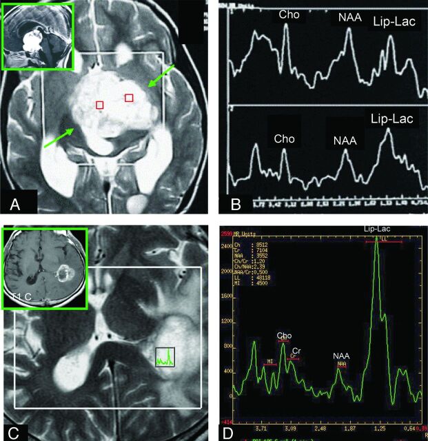
Fig 3.
Comparison of the Lip-Lactu/Crtu ratio on short-TE multivoxel MR spectroscopy in PA and HGA. A and C, Axial T2-weighted imaging was used for positioning of the regions of interest in the tumor. A and B, The solid homogeneous enhancing hypothalamic-chiasmatic PA has a Lip-Lac peak. The (C and D) necrotic ring-enhancing GBM located on the lateral wall of the left lateral ventricular trigone also has an elevated Lip-Lac peak (D), but the Lip-Lactu/Crtu ratio in this tumor is higher than that found in the PA (B). A and B reproduced with permission from Aragao et al.47

