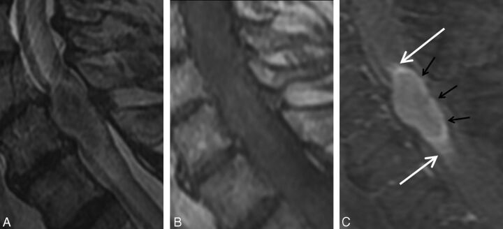
Fig. 3.
Concurrent rim and flame signs. A 68-year-old man with metastatic non-small cell lung carcinoma who presented with progressive bilateral lower extremity weakness. MR imaging of the cervical spine demonstrates an intramedullary spinal cord metastasis at C6. Sagittal T2-weighted (A), T1-weighted (B), and postcontrast T1-weighted fat-suppressed (C) images. Note the enhancing intramedullary mass with a thin rim of more intense enhancement (rim sign; black arrows, C) as well as the ill-defined flame-shaped regions of enhancement at the superior and inferior margins of the mass (flame sign; white arrows, C).

