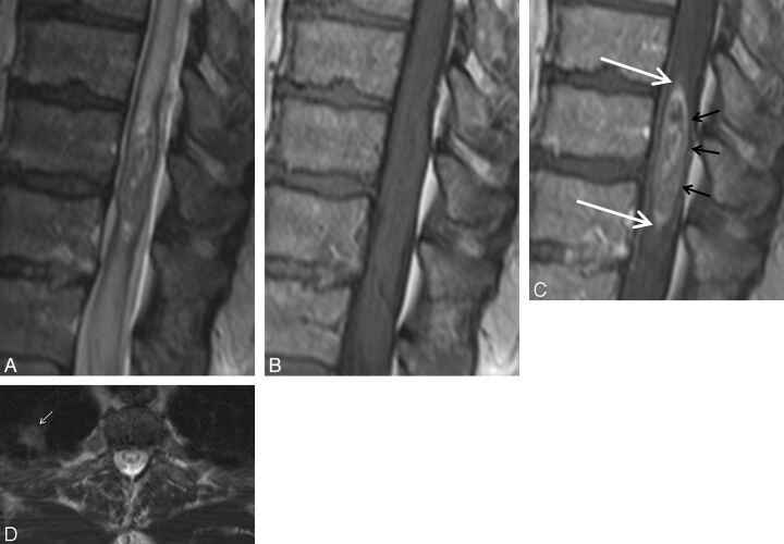
Fig. 4.
Pathologically proved intramedullary spinal cord metastasis demonstrating the potential utility of the rim and flame signs. A 55-year-old man who presented with 8 months of progressive lower extremity paresthesias. MR imaging of the thoracic spine demonstrates an intramedullary spinal cord metastasis due to small cell lung carcinoma. Sagittal T2-weighted (A), T1-weighted (B), postcontrast T1-weighted (C), and axial T2-weighted (D) images. Note the concurrent rim (black arrows, C) and flame signs (white arrows, C). Despite these findings and the observation of a small right upper lobe mass (small white arrow, D), the radiologist's primary consideration was ependymoma. The clinicians agreed with this impression. Bronchoscopic biopsy of mediastinal adenopathy yielded small cell carcinoma. Because of the impression of the cord mass being an ependymoma, a diagnostic partial resection was performed, yielding metastatic small cell carcinoma. Knowledge of the rim and flame signs as specific findings of ISCM could potentially have avoided diagnostic biopsy.

