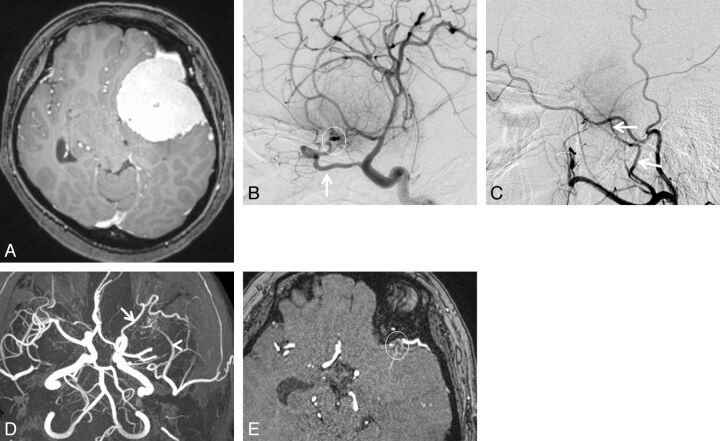
Fig. 1.
A 43-year-old woman with sphenoid ridge meningioma. A, Axial contrast-enhanced 3D turbo field echo image showing a large enhanced mass in the left anterior to middle cranial fossa regions. B, DSA (lateral projection from the left internal carotid artery) reveals a tumor fed primarily by the recurrent meningeal artery of the ophthalmic artery (arrow). Based on the surgical findings, the dural attachment was the sphenoid ridge (circle). C, DSA (lateral projection from the left external carotid artery) shows a tumor fed partially by the middle meningeal artery (arrows). The right middle meningeal artery was judged to be the secondary feeder. D, 3D TOF MRA (axial projection) depicts dilated branches from the left ophthalmic (arrow) and middle meningeal arteries (arrowhead). E, This axial source 3D TOF MRA image shows tumor-feeding branches from the left ophthalmic artery at the left sphenoid ridge (circle). Both readers judged that the ophthalmic artery was the primary feeder and that the dural attachment was the left sphenoid ridge.

