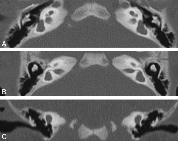
Fig 4.
Patient M. CT. Axial views (A and B) through the cochlea and the vestibule show a small cochlea, flattened with a partition hardly visible and atresia of cochlear nerve canals, an enlarged vestibular cavity, and agenesis of all of the semicircular canals. Coronal view (C) confirms the absence of the superior and lateral SCC. Posterior deformity of the vestibule cannot exclude an anlage.

