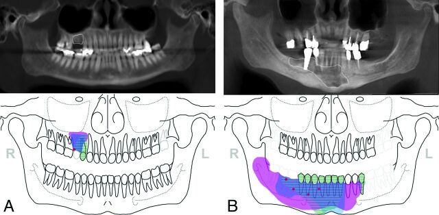
Fig 2.
Location and extent of BONJ as marked on jaw schemes from contrast-enhanced MR imaging (blue), [18F] fluoride PET/CT (pink), clinical preoperative (red), and intraoperative examinations (striated areas) were graphically assembled in 1 image and related to delineations (white contours) on panoramic views from CBCT to compare the extent of BONJ among different modalities/examinations. A, In patient 9, only a small clinically suspicious area in the right maxilla was seen. PET/CT and CEMR imaging showed slightly larger areas of suspected BONJ than CBCT and intraoperative examinations in the right maxilla (regions 14–15). BONJ was histologically proved. B, In patient 2, BONJ was suspected preoperatively in the regions 44–46 of the right mandible. The actual extent of the BONJ was markedly larger than expected clinically and on CBCT in both CEMR imaging and PET/CT. This was confirmed intraoperatively, where only parts of the necrotic bone were removed.

