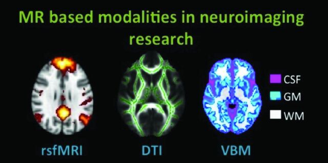
Fig 1.
MR imaging−based modalities in neuroimaging research discussed in this review. In rsfMRI, the DMN of healthy subjects is shown. In DTI, the skeleton (green) is overlaid on the average FA image of healthy subjects. In VBM of healthy subjects, GM (blue-light blue), WM (white), and CSF (pink) can be extracted from T1-weighted structural data. All images are schematic representations of our own data, processed with Functional MR Imaging of the Brain Software Library, Version 1.7 (http://www.fmrib.ox.ac.uk).

