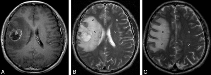
Fig 4.
Solitary MET with peritumoral T2 prolongation of grade II. Postcontrast axial T1WI shows a ringlike enhancing lesion in the right frontal lobe with a maximal diameter of 3.6 cm (A). On the same section as A, axial T2WI demonstrates strong peritumoral T2 prolongation, which is larger in area than the tumor (B). On this section, 2 ROIs and 2 mirror ROIs are placed in the prolonged region (black ring) and contralateral area (white ring). On the section superior to A, another 2 ROIs with 2 mirror ROIs are positioned in the prolonged region and the contralateral region, respectively (C). The nSI is 2.75 (603.5/219.5).

