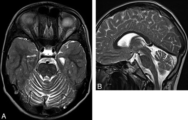
Fig 2.
Ataxia-telangiectasia. A, Axial T2 image shows cortical thinning and fissure enlargement in the vermis and cerebellar hemispheres. B, Midline sagittal T2 image demonstrates an atrophic vermis but normal volume of the brain stem. Although nonspecific, this pattern of diffuse cerebellar atrophy suggests a degenerative disease.

