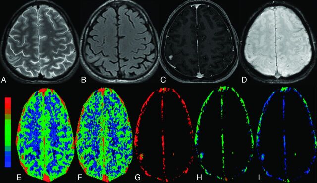
Fig 2.
The granular-nodular stage of NCC with minimal edema in the right parietal region in a 32-year-old man who presented with recurrent partial seizures. The cyst appears hypointense on T2 image (A) and FLAIR image (B) with hyperintense minimal perifocal edema. C, The lesion shows nodular enhancement on a postcontrast T1-weighted image. D, On SWAN image, the lesion shows some hypointensity, which suggests minimal mineralization. On color-coded CBF [rCBF = 3.5 (E)], CBV [rCBV = 2.4 (F)], kep [kep = 1.1 minute−1 (G)], Ktrans [Ktrans = 0.15 minutes−1 (H)], and ve [ve = 0.16(I)] maps, the abnormal changes are clearly evident in and around the lesion.

