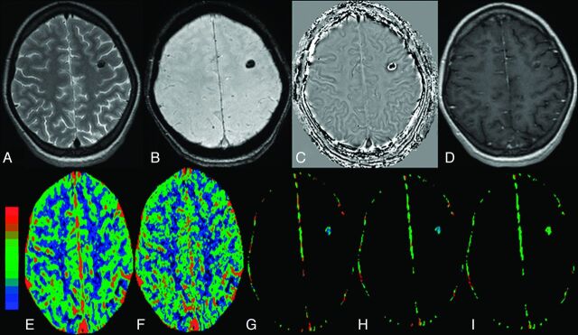
Fig 3.
The calcified stage of NCC without edema in the left frontal lobe in a 34-year-old woman who presented with recurrent complex partial seizures. The lesion appears hypointense on T2 image (A) and SWAN image (B) and shows a negative phase in the center with a peripheral positive phase consistent with calcified cysticercous granuloma on a filtered phase image extracted from the SWAN imaging (C). Postcontrast T1-weighted image does not show any definite enhancement (D). On color-coded CBF [rCBF = 1.5 (E)], CBV [rCBV=1.3 (F)], kep [kep = 0.8 minutes−1 (G)], Ktrans [Ktrans = 0.02 minutes−1 (H)], and ve [ve = 0.05 (I)] maps, the abnormal changes are clearly evident in and around the lesion.

