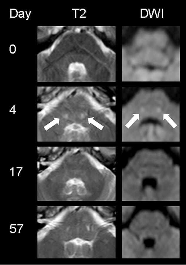
Fig 2.
MR imaging findings of subject 2 presenting with aphasia, apraxia, and memory deficits. At the level of the fourth ventricle, symmetric round signal hyperintensities (filled white arrows) can be visualized in the central pons, normalizing with clinical improvement at day 17. (The DWI image on day 4 has been modified from Reference 22.)

