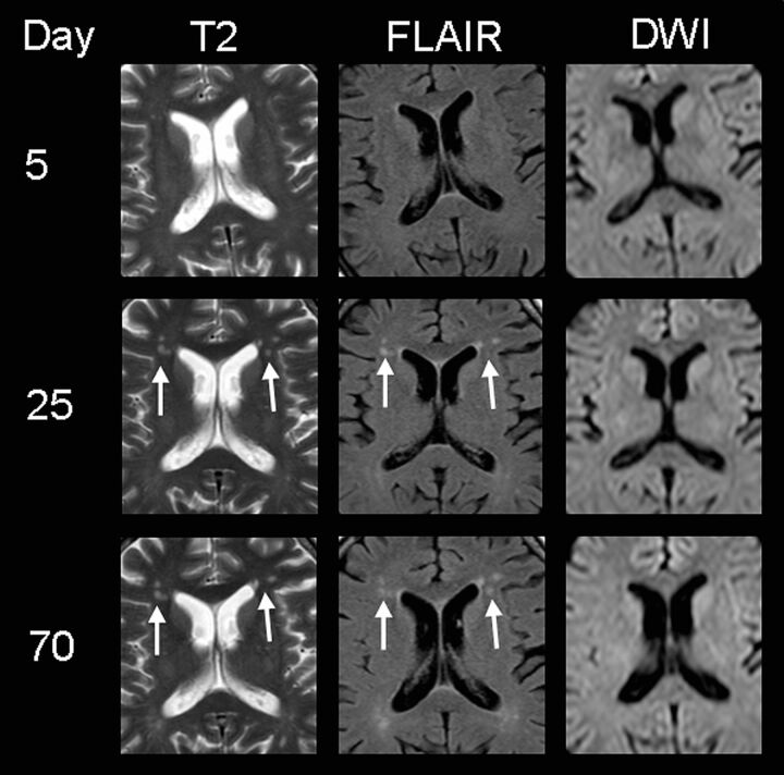
Fig 3.
MR imaging findings of subject 3, presenting with delirium and memory deficits. Note the patchy hyperintensities on T2WI and FLAIR images in the frontal periventricular white matter (white arrows) 3 weeks after the onset of neurologic symptoms (supposedly as a result of HUS encephalopathy because they could not be detected on day 5). DWI shows no restriction, indicating that no ischemia is present. At this stage, the patient did not show any neurologic symptoms. After 10 weeks, these lesions are still visible despite complete clinical recovery.

