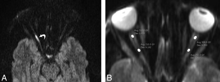
Fig 1.
A 37-year-old man with right-sided TON following a motor vehicle collision. Axial diffusion-weighted image (A) shows hyperintensity of the posterior segment of the optic nerve (curved arrow) due to restricted diffusion. B, Axial DWI (b=0) of the same patient shows ROI placement on the anterior and posterior segments of intraorbital optic nerve.

