
Fig 2.
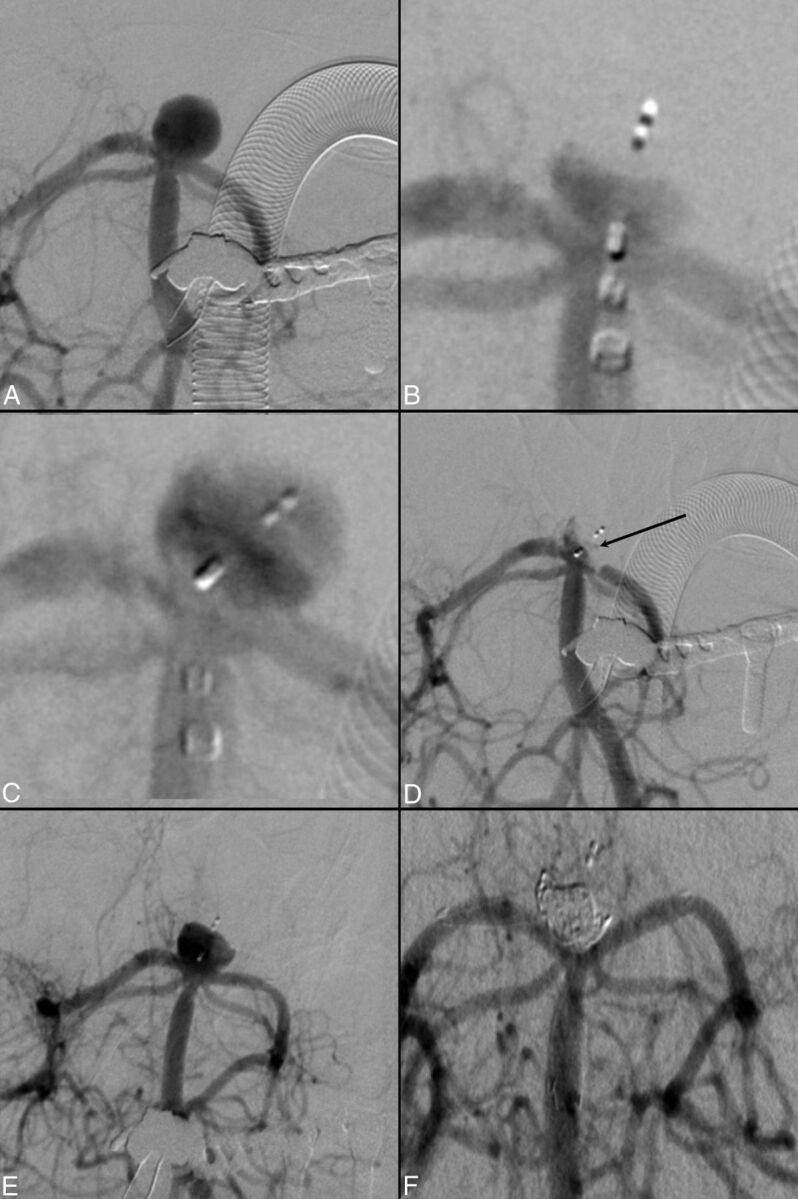
Unruptured basilar artery tip aneurysm and posterior cerebral artery stenosis after migration of the WEB and retreatment with stent and coils. A, Initial angiogram of the aneurysm. B, Deployment of the WEB-DL 7 × 4 mm. C, Migration of the device toward the left side of the aneurysm. D, High-grade stenosis of the left posterior cerebral artery P1 segment (arrow), which was stented subsequently with a Neuroform stent (Stryker Neurovascular, Fremont, California). E, Control angiogram at 3 months shows an aneurysm remnant. F, Aneurysm occlusion after Y-stent placement (Low-Profile Visualized Intraluminal Support Device; MicroVention, Tustin, California) and additional coil embolization.