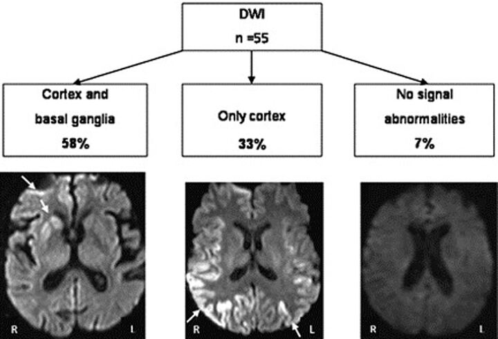
Fig 1.
DWI in CJD. The most frequent MR imaging lesion patterns were defined by using DWI as the most sensitive technique. Cortex and basal ganglia hyperintensity was observed in approximately two-thirds (58%), and isolated cortical hyperintensity, in one-third (33%).7

