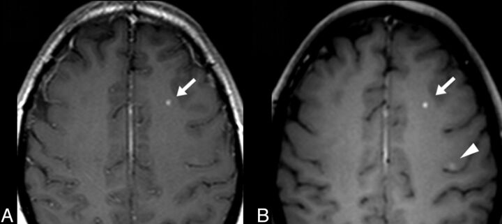
Fig 2.
Postcontrast T1-weighted 2D SE (A) and 4-mm-thick axial reformatted views of the 3D TSE (B) images of patient 24 with MS. An active enhanced MS lesion is detected within the left frontal white matter by using both axial 2D SE images (A, arrow) and a 4-mm-thick axial reformatted view of the 3D TSE sequence (B, arrow). Due to improved image contrast, an additional lesion involving the precentral sulcus is visible by using the 3D TSE sequence (B, arrowhead).

