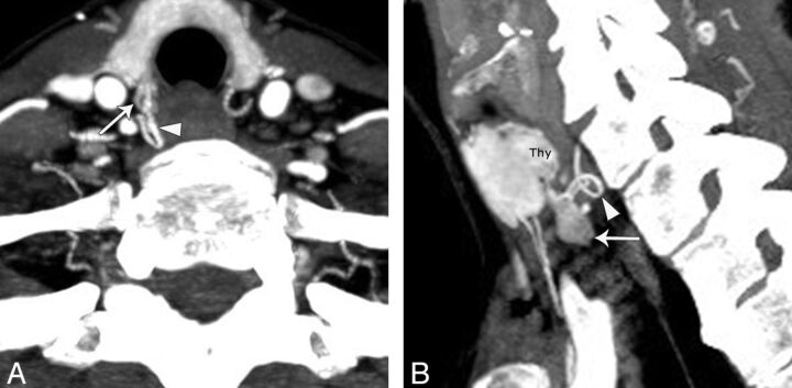
Fig 1.
A 69-year-old woman with a right parathyroid adenoma. Axial (A) and sagittal (B) arterial phase images demonstrate an oval-shaped hyperenhancing adenoma posterior and inferior to the lower pole of the right thyroid gland (straight arrows). A characteristic tortuous feeding artery is seen at the superior aspect of the adenoma (arrowheads).

