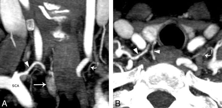
Fig 3.
A 70-year-old woman with a right parathyroid adenoma. A, Coronal arterial phase image demonstrates an oval hyperenhancing adenoma in the right tracheoesophageal groove (straight arrow). Coronal (A) and axial (B) images show an enlarged inferior thyroid artery (arrowheads) arising from the right subclavian artery and terminating at the superior pole of the adenoma. Note the normal contralateral inferior thyroid artery (curved arrows).

